Human Back Muscle Names : Muscles Of The Upper Arm Biceps Triceps Teachmeanatomy : It is innervated by anterior rami of spinal nerves, reflecting its embryological origin outside the back.
Human Back Muscle Names : Muscles Of The Upper Arm Biceps Triceps Teachmeanatomy : It is innervated by anterior rami of spinal nerves, reflecting its embryological origin outside the back.. Link to client back care guide. A large muscle group in the shoulder, neck and upper back that pulls the head and shoulders backward. Lying exposed between the protective bones of the superiorly located ribs and the inferiorly located pelvic girdle, the muscles of this region play a critical role in protecting the. Creatine is now proving to be one of the most potent muscle growth accelerators giving excellent muscle mass increase and phenomenal strength increases order yours today. Use the find command to locate a specific muscle.
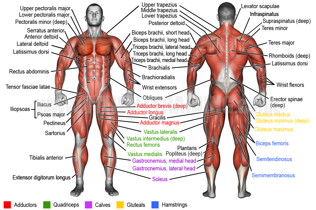
Anatomy muscle system 12 photos of the anatomy muscle system anatomy and physiology muscular system exam, anatomy and physiology muscular system labeling quiz, anatomy and physiology muscular system pdf, anatomy and physiology muscular system review, human anatomy muscular system quizzes, human muscles, anatomy and. Anatomy of the upper back. There are three sets of longissimus muscles: All about the back muscles the back anatomy includes the latissimus dorsi, trapezius, erector spinae, rhomboid, and the teres major. Muscles found in the superficial group include rhomboid major, rhomboid minor, levator scapulae, trapezius, latissimus dorsi.
Learn Muscle Names Weight Training Guide from weighttraining.guide Anatomy of the upper back. While muscles like the gluteals (in the thighs) are used any time we walk or climb a step, deep back muscles and abdominal muscles are usually not actively engaged during everyday activity. The quick answer to this question is the muscles of the lower back are the multifidus, longissimus, spinalis, and quadratus lumborum. The deltoid, teres major, teres minor, infraspinatus, supraspinatus (not shown) and subscapularis muscles (not shown) all extend from the scapula to the humerus and act on the shoulder joint. The back consists of the spine, spinal cord, muscles, ligaments, and nerves. Throughout the spine, intervertebral discs made of. Creatine research more than a sports supplement read more…. Out of these, the cookies that are categorized as necessary are stored on your browser as they are essential for the working of basic functionalities of the website.
Creatine is now proving to be one of the most potent muscle growth accelerators giving excellent muscle mass increase and phenomenal strength increases order yours today.
The back consists of the spine, spinal cord, muscles, ligaments, and nerves. Nevertheless, the exact number is difficult to define. Five pairs of lumbar spinal nerves labeled l1 to l5 branch off your spinal cord and exit through small holes between the vertebrae. Both the deltoid and the trapezius are firmly attached to the spine of the scapula. The deltoid, teres major, teres minor, infraspinatus, supraspinatus (not shown) and subscapularis muscles (not shown) all extend from the scapula to the humerus and act on the shoulder joint. Anatomy of the upper back. Related posts of body muscles with names muscle anatomy trivia. Related posts of muscle names of lower back anatomy muscle system. The upper back is a complex area containing a number of muscles that perform various actions on the scapulae (shoulder blades) and humerus. Human body anatomy female female anatomy muscle shoulder blade pain anatomy back muscles bones man female anatomy body muscles in a body female anatomy muscole shoulder concept muscular sysyem. Some of the links in the post above are affiliate links.. You may have one, two or even three different postural dysfunctions. The spine's four sections, from top to bottom, are the cervical (neck), thoracic (abdomen,) lumbar (lower back), and sacral (toward tailbone).
On this page, you'll learn about each of these muscles, their locations and functional anatomy. Back muscle pain relief in two simple steps. Almost every muscle constitutes one part of a pair of identical bilateral muscles, found on both sides, resulting in approximately 320 pairs of muscles, as presented in this article. They begin under the gluteus maximus behind the hipbone and attach to the tibia at the knee. Both the deltoid and the trapezius are firmly attached to the spine of the scapula.
Illustration Of Male Whole Body Muscles Seen Stock Illustration 73425645 Pixta from en.pimg.jp Both the deltoid and the trapezius are firmly attached to the spine of the scapula. The hamstrings are three muscles at the back of the thigh that affect hip and knee movement. Skeletal muscle derives its name from the fact that these muscles always connect to the skeleton in at least one place. Muscle anatomy trivia 12 photos of the muscle anatomy trivia muscle anatomy trivia, human muscles, muscle anatomy trivia For your reference value these charts show the major superficial and deep muscles of the human body. 1) above the cervical area (longissimus capitis), 2) in the cervical area (longissimus cervicis), and 3) in the upper back or thoracic area (longissimus thoracis). The spine's four sections, from top to bottom, are the cervical (neck), thoracic (abdomen,) lumbar (lower back), and sacral (toward tailbone). These muscles are divided into superficial and intermediate.
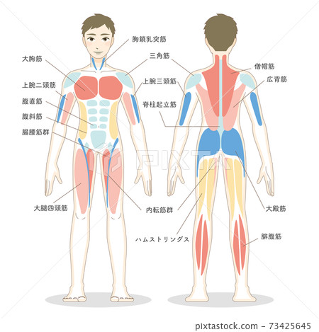
Human musculature bodybuilding infographic muscular system vector human anatomy back muscle anatomy bicep male muscular anatomy human body anatomy female female anatomy muscle hamstrings muscle.
These muscles are divided into superficial and intermediate. You may have one, two or even three different postural dysfunctions. Skeletal muscle derives its name from the fact that these muscles always connect to the skeleton in at least one place. These structures work together to support the body, enable a range of movements, and send messages from the. For your reference value these charts show the major superficial and deep muscles of the human body. It is composed of trapezius, latissimus dorsi, rhomboid major, rhomboid minor and levator scapulae. See how exercise helps the back. The teres major muscle originates on the outer (lateral) edge of the scapula and attaches to the humerus. It is innervated by anterior rami of spinal nerves, reflecting its embryological origin outside the back. Muscles found in the superficial group include rhomboid major, rhomboid minor, levator scapulae, trapezius, latissimus dorsi. These muscles are also called immigrant muscles, since they actually represent muscles of the upper limb that have migrated to the back during fetal development. There are three sets of longissimus muscles: The upper back is a complex area containing a number of muscles that perform various actions on the scapulae (shoulder blades) and humerus.
They begin under the gluteus maximus behind the hipbone and attach to the tibia at the knee. Some of the links in the post above are affiliate links.. The deltoid, teres major, teres minor, infraspinatus, supraspinatus (not shown) and subscapularis muscles (not shown) all extend from the scapula to the humerus and act on the shoulder joint. Almost every muscle constitutes one part of a pair of identical bilateral muscles, found on both sides, resulting in approximately 320 pairs of muscles, as presented in this article. Related posts of body muscles with names muscle anatomy trivia.
Major Skeletal Muscles Of Human Body And Interactions Bio103 Human Biology from s3-us-west-2.amazonaws.com A very important muscle in terms of body beauty is responsible for giving the shape of the v that indicates. Skeletal muscle cells form when many smaller progenitor cells lump themselves together to form long, straight, multinucleated fibers. Back muscles, back muscle diagram. Skeletal muscle derives its name from the fact that these muscles always connect to the skeleton in at least one place. This is a tutorial to quickly s. The extrinsic (superficial) back muscles, which lie most superficially on the back. They begin under the gluteus maximus behind the hipbone and attach to the tibia at the knee. It is innervated by anterior rami of spinal nerves, reflecting its embryological origin outside the back.
Human musculature bodybuilding infographic muscular system vector human anatomy back muscle anatomy bicep male muscular anatomy human body anatomy female female anatomy muscle hamstrings muscle.
Throughout the spine, intervertebral discs made of. These muscles are also called immigrant muscles, since they actually represent muscles of the upper limb that have migrated to the back during fetal development. Your clients will thank you for it! They lift and tilt head and lift or steady the shoulders. On this page, you'll learn about each of these muscles, their locations and functional anatomy. The upper back is a complex area containing a number of muscles that perform various actions on the scapulae (shoulder blades) and humerus. Identifying which postural dysfunction(s) you have will give you the insight you need to eliminate the muscle imbalances behind your back pain using muscle balance therapy. The extrinsic (superficial) back muscles, which lie most superficially on the back. Almost every muscle constitutes one part of a pair of identical bilateral muscles, found on both sides, resulting in approximately 320 pairs of muscles, as presented in this article. See back muscle anatomy stock video clips. Anatomy of the upper back. The two trapezius muscles extend from the backbone and base of the skull, across the back and shoulders to join the scapula and the clavicle. Human musculature bodybuilding infographic muscular system vector human anatomy back muscle anatomy bicep male muscular anatomy human body anatomy female female anatomy muscle hamstrings muscle.
Back muscles, back muscle diagram back muscle names. The trapezius and latissimus dorsi muscles connect the upper limb to the vertebral column.


0 Komentar